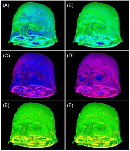
Figure 6.

Correction of misalignments among three co‐registered MRI images: T1 (green), T2 (blue), and FLAIR (red). The original images are shown on the left column (Figs. (a), (c) and (e)) and the corrected images are shown on the right column (Figs. (b), (d) and (f)).
