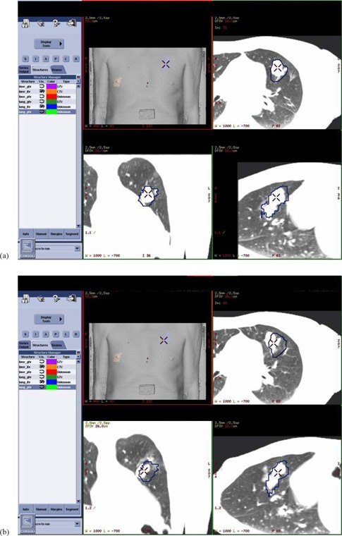
Figure 1.

4DCT images: (a) at full exhale respiratory phase on simulation day, and (b) at full exhale respiratory phase on treatment day #4 for one patient (case #6) in this study. The contoured ITV contained the tumor motion envelope on the day of simulation, but the tumor can be seen to have shifted for treatment day #4. Image guidance shifts were carried out to align to the target's day #4 location prior to treatment delivery. In this example, the corrective shifts were 2 mm posteriorly and 9 mm to patient left.
