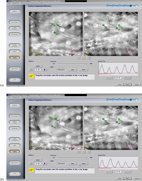
Figure 3.

Two orthogonal X‐ray images acquired at the full exhale reference level of a liver tumor patient with the implanted fiducial markers before (a) and after (b) fusion. Markers (M1, M2, and M3) were reconstructed from planning CT of the location of the implanted fiducial markers and then were projected onto the X‐ray images.
