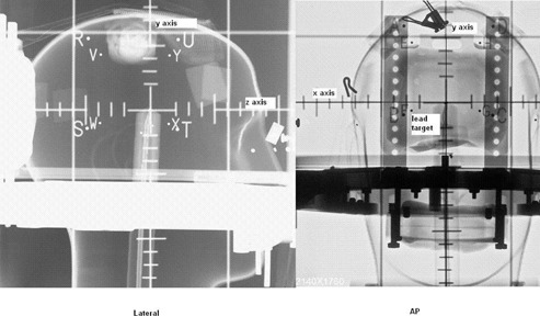
Figure 2.

Computed radiographic film of the head phantom fitted with GTC frame and the angiographic localizer box. Small lead target is placed at the center Lateral (left) and AP (right) images are taken and mismatch of the target compared to the fiducials are found. The direction of x, y and z axes are also illustrated.
