Figure 6.

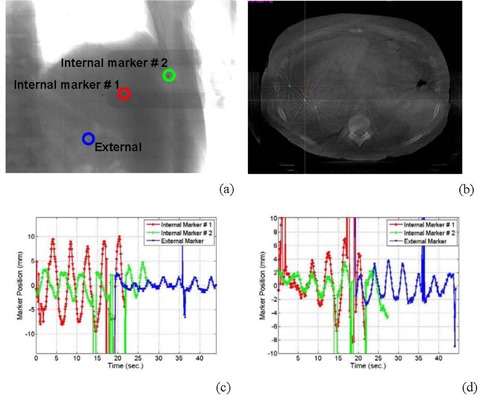
Internal and external seed markers in a radiographic projection (a) and a CBCT image (b); the internal (c) and external (d) markers’ motion shown in (a) and (b) parallel and perpendicular to the superior‐inferior, respectively.

Internal and external seed markers in a radiographic projection (a) and a CBCT image (b); the internal (c) and external (d) markers’ motion shown in (a) and (b) parallel and perpendicular to the superior‐inferior, respectively.