Figure 4.

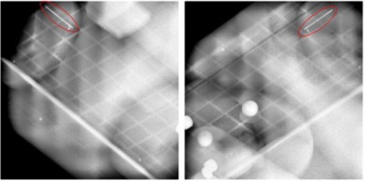
A pair of orthogonal X‐ray images taken with the ExacTrac system. The implanted coil (circled in red) is seen at the top left in the left image and at the top right in the right image.

A pair of orthogonal X‐ray images taken with the ExacTrac system. The implanted coil (circled in red) is seen at the top left in the left image and at the top right in the right image.