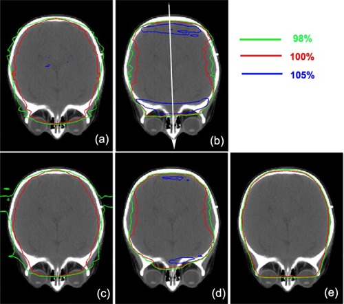
Figure 2.

2D dose distributions near the subfrontal cribriform plate level in an axial view for patient #1 (listed in Table 1). Proton plans of the Eclipse and the XiO TPS with and without using compensator are shown: (a) Eclipse without compensator, (b) Eclipse with compensator, (c) XiO without compensator, and (d) XiO with compensator. A photon plan of the Pinnacle 3 TPS is shown in panel (e). Isodose lines of 105%, 100% and 98% with respect to prescription dose (23.4 Gy (RBE)) are shown.
