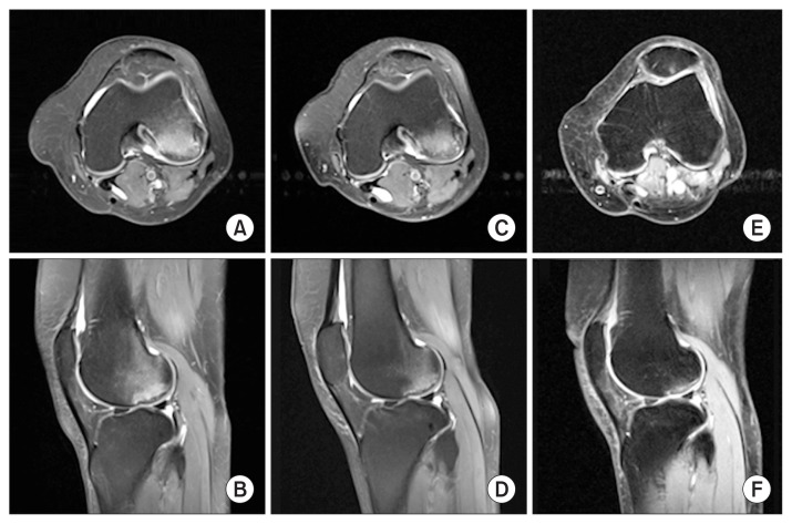
Fig. 2.
Axial (top) and sagittal (bottom) magnetic resonance imaging scans demonstrating the progression of the patient’s subchondral insufficiency fracture at presentation (A, B) and at 6 months (C, D) and 30 months of follow-up (E, F). Note the decreasing area of bone marrow oedema in the femoral condyle. There is significant articular oedema at 30 months as the image was taken on the day immediately after the patient had run a marathon.

