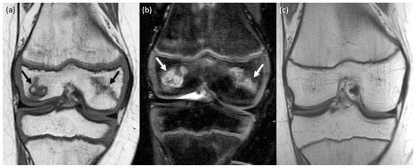
Fig. 2.
Spontaneous resolution of ON in a 10-year old girl. (a) Coronal T1-weighted image (TR/TE/Flip angle: 516/17/90) and (b) coronal T2-weighted fat saturated Fast Spin-Echo image (TR/TE/Flip angle: 3700/61/90) shows ON in the medial and lateral epiphysis of the distal femur (arrows). (c) Follow up MRI 3 years later reveals complete resolution of the osteonecrosis in the epiphysis of the distal femur.

