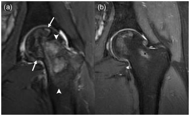
Fig. 3.
Nearly complete resolution of ON after decompression surgery in a 20-year old boy at diagnosis. (a) Coronal T2-weighted fat saturated Fast Spin-Echo image (TR/TE/Flip angle: 4356/49/111) showing large ON in the left femoral head (arrows) with extensive bone marrow oedema (arrowheads) in the adjacent head and neck and (b) coronal PD fs image (TR/TE/Flip angle: 2798/35/111) with only minimal residual ON in femoral head after decompression surgery. Of note, partially shown postprocedural tract (*).

