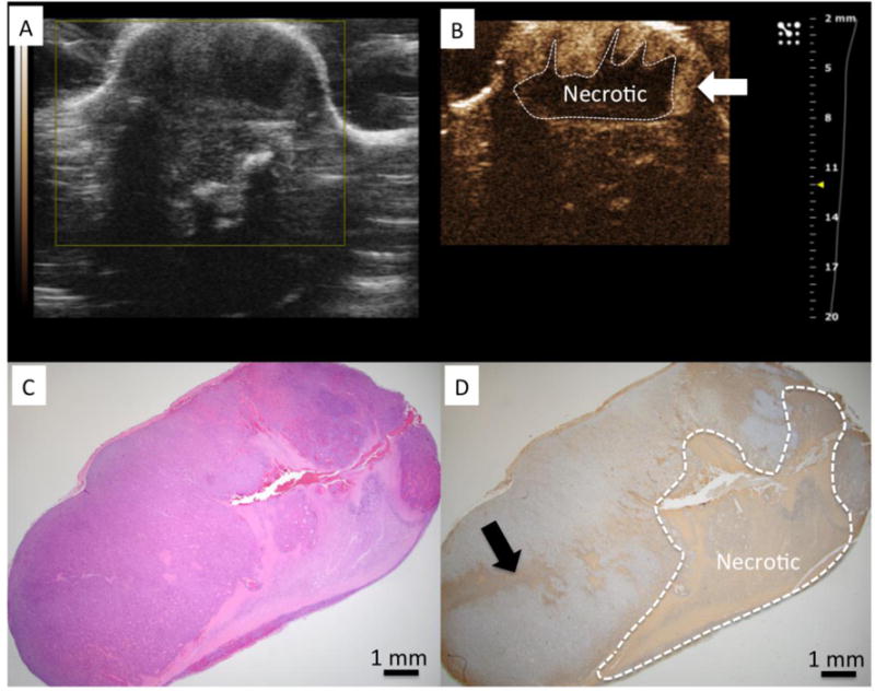
Figure 2.

A) US imaging shows tumor to have heterogeneous echo properties. B) CEUS identified a central necrotic area within the tumor after VTP (dashed line) C) H&E stained slide of the tumor corresponding to US imaging plane. D) TUNEL demonstrates a region of positive (brown) staining clearly demarcating a region of necrosis similar in size and shape to region of non-enhancing region observed on CEUS. Regions of enhancement on CEUS correspond to regions without positive staining for TUNEL and was interpreted to be viable tissue on H&E.
