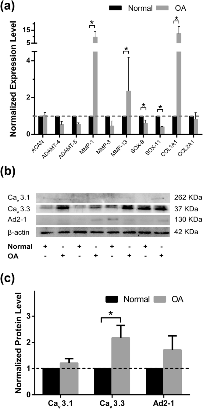
Figure 5.

Expression level of chondrogenic genes and protein abundance of T-VDCCs in non-degenerated and early-degenerated articular cartilage samples. (a) Compared with non-degenerated cartilage (Normal, black bars), chondrocytes located in early-degenerated area (OA, grey bars) of TKA samples showed increased expression level of MMP-1, MMP-13, and COL1A1; and decreased SOX-9 and SOX-11 (* P < 0.05) (n = 6). (b) Representative Western blot detection of Cav3.1, Cav3.3, A2D-1, and β-actin (full-length blots/gels were presented in Supplementary Figure S1). (c) Compared with non-degenerated cartilage (Normal, black bars), significantly increased protein level of Cav3.3 was noticed in early-degenerated (OA, grey bars) regions (* P < 0.05) (n = 8).
