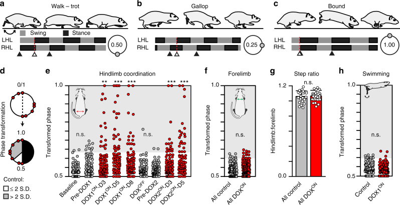
Fig. 4.
Silencing L2–L5 interneurons selectively disrupts left–right hindlimb alternation during overground stepping. a–c Stereotypic locomotor gaits with representative swing–stance graphs and characteristic left–right hindlimb coordination values (shown in circular plots, see Methods for detail). d Schematic illustrating phase transformation (see Methods for detail). Shaded area denotes any coordination value beyond normal variability observed (>2 S.D.) at control time points. Each circle represents one step cycle (n = 84/time point; n = 10–14/animal). e Silencing L2–L5 interneurons significantly increased the proportion of steps that deviated beyond normal variability observed at control time points. Removing DOX restored alternation and silencing one month later repeated the effects (Baseline, n = 3/84 vs. DOX1ON, n = 15/84 (p = 0.002, z = 3.08), 24/84 (p < 0.001, z = 4.69), 17/84 (p = 0.001, z = 3.45); Pre-DOX2, n = 2/84 vs. DOX2ON, n = 15/84 (p = 0.001, z = 3.44), 21/84 (p < 0.001, z = 4.52); **p ≤ 0.01, ***p ≤ 0.001; Binomial Proportion (B.P.) Test). Control time points were not significantly different from each other (Baseline vs. Pre-DOX1, p = 0.116, z = 1.57; DOXOFF vs. Pre-DOX2, p = 0.562, z = 0.58). f Left–right forelimb alternation was not perturbed (All control vs. All DOXON, p = 0.322, z = 0.99). g No differences were observed in the hindlimb/forelimb stepping index (circles = individual animal’s average hindlimb:forelimb step ratio at each time point; Control: N = 24 total average step ratios; DOX: N = 30 total average step ratios; bars represent group average ± S.D.; p = 0.75, critical t = 2.003, df = 52, two-sample t-test). h Left–right hindlimb alternation persisted during swimming (n = 80 stroke cycles/time point; p = 0.20, z = 1.28, B.P. test). In a-c, classic gait data were generated from a separate study, see Methods for detail. Illustrations by A. Pocratsky

