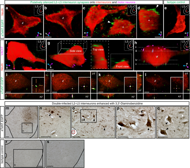
Fig. 7.
Immunohistochemical detection of eTeNT.EGFP in the double-infected L2–L5 interneurons. a–d Volumetric three-dimensional renderings show eTeNT.EGFP+ terminals closely apposed to somata and primary dendrites of neurons in the intermediate gray matter (#,* indicate same neuron rotated, x–y–z axes shown in gray/magenta/blue). e Isotype control showed little to no signal (nonimmune rabbit sera at 5 mg/ml, 1:5000 dilution). f–h Volume rendered images show eTeNT.EGFP+ terminals surrounding motor neurons marked by vesicular acetylcholine transferase (VAChT, magenta) in the caudal lumbar segments. Cross-sections throughout the z-stack confirm colocalization of VAChT with eTeNT.EGFP in the x–z and y–z planes (i–l, white signal in crosshairs; cross-sections throughout motor neuron shown in (f); a–f scale = 10 µm). Amplification of eTeNT.EGFP signal with 3,3′-Diaminobenzidine (DAB) revealed double-infected neurons within the intermediate gray matter of the L2 spinal segment (m–q dark arrowheads = eTeNT.EGFP-positive neurons; white arrowheads = eTeNT.EGFP-negative neurons). r, s Isotype controls revealed no DAB-enhanced eTeNT.EGFP signal. m–o, r, s scale = 100 µm; p, q scale = 50 µm

