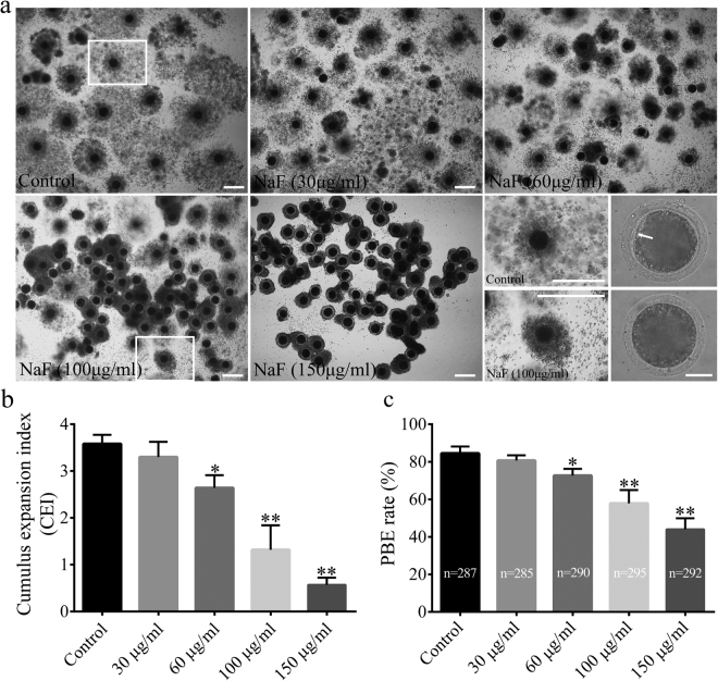
Figure 1.
Effect of NaF exposure on cumulus cell expansion and PBE rate in porcine oocytes. (a) COCs were cultured with 30, 60, 100, and 150 μg/ml NaF for 44 h. Cumulus cell expansion decreased after NaF exposure; COCs: scale bar = 200 μm; oocyte: scale bar = 50 μm. (b) Average CEI was calculated at 44 h for COCs in each experimental group. (c) PBE rate decreased significantly after NaF treatment. Data are presented as mean ± SD of three independent experiments. Statistically significant differences are indicated by asterisks (*p < 0.05 and **p < 0.01).

