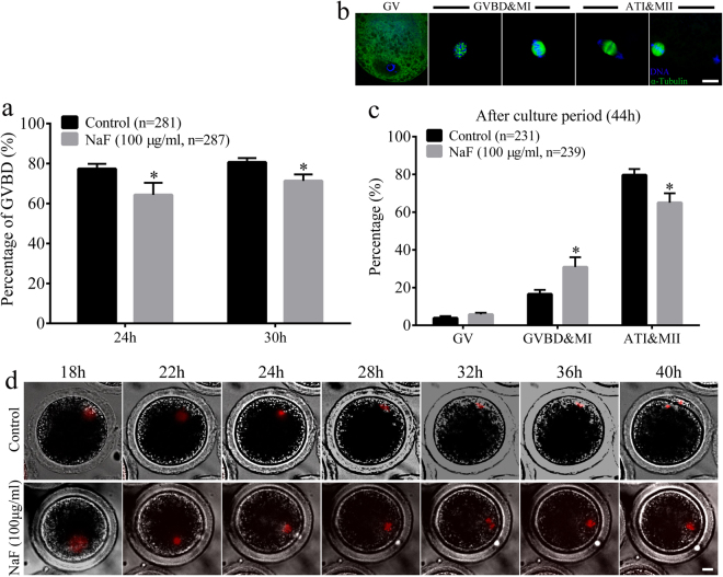
Figure 3.
NaF exposure affects cell cycle progression in porcine oocyte. (a) The percentage of oocytes undergoing GVBD after NaF treatment for 24 or 30 h. (b) Representative confocal images of porcine oocytes in various maturation phases; scale bar = 10 μm; blue, DNA; green, α-tubulin. (c) Percentages of cells in various meiotic maturation phases were calculated at 44 h. (d) Time-lapse images obtained after microinjecting GV-stage oocytes with histone H2B–mCherry cRNA; Scale bar = 20 μm. ATI: anaphase/telophase I; GV: germinal vesicle; GVBD: germinal vesicle breakdown; MII: metaphase II. Data are presented as mean ± SD of three independent experiments. Statistically significant differences are indicated by asterisks (*p < 0.05).

