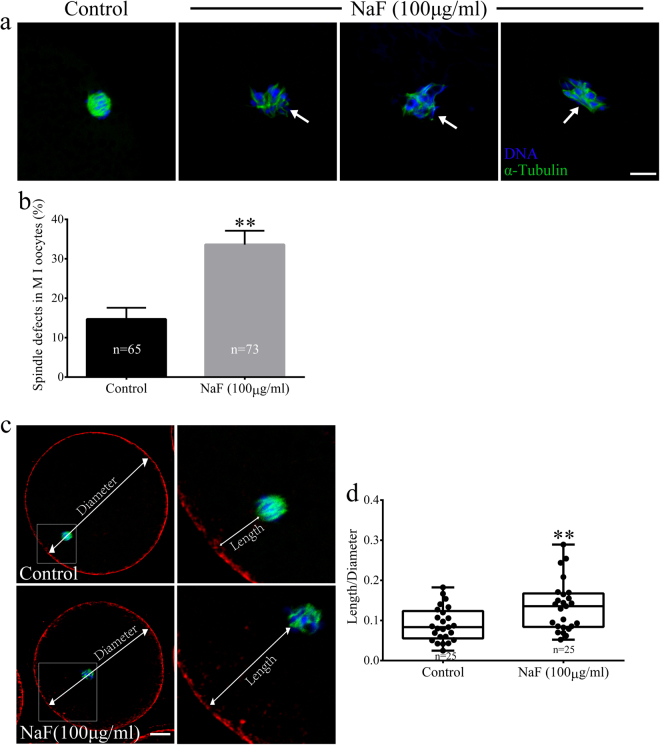
Figure 4.
NaF exposure induces defects in spindle assembly in porcine oocytes. (a) Representative spindle morphologies of oocytes in the MI stage. Control oocytes showed normal spindle morphology, whereas NaF-treated oocytes showed various spindle defects (arrows); scale bar = 10 μm; blue, DNA; green, α-tubulin. (b) Percentages of NaF-treated oocytes in the MI stage showing aberrant spindle morphology. (c) Spindle positioning in oocytes in the MI stage. Spindles in control oocytes were located almost peripherally, whereas those in NaF-treated oocytes were located almost centrally; scale bar = 20 μm. (d) Quantification of the distance between the spindle and cell cortex (length/diameter) in control and NaF-treated oocytes. Data are presented as mean ± SD of three independent experiments. Statistically significant differences are indicated by asterisks (**p < 0.01).

