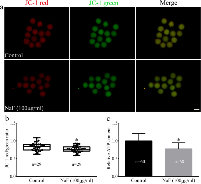
Figure 6.
NaF exposure induces mitochondrial dysfunction (as determined by measuring ΔΨm and ATP production) in porcine oocytes. (a) JC-1 staining of NaF–treated oocytes. ΔΨm was significantly lower in NaF-treated oocytes than in control oocytes; scale bar = 100 μm. (b) Fluorescence intensity of JC-1 in oocytes. (c) Intracellular ATP content in oocytes. NaF exposure clearly affected mitochondrial ATP production. Data are presented as mean ± SD of three independent experiments. Statistically significant differences are indicated by asterisks (*p < 0.05).

