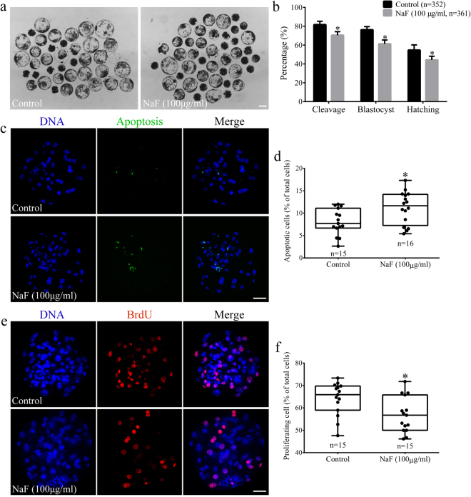
Figure 9.
NaF exposure impairs the further developmental potential of porcine oocytes. (a) NaF exposure decreased the developmental potency of oocytes after parthenogenetic activation; scale bar = 100 μm. (b) Embryonic development rates of control and NaF-treated oocytes. (c) Representative images of embryos in the blastocyst stage after performing the TUNEL assay; scale bar = 50 μm. (d) Percentages of apoptotic cells in blastocysts developed in vitro. (e) Immunofluorescent staining of BrdU in blastocysts; scale bar = 50 μm. (f) Percentages of BrdU-positive cells in the blastocysts. Data are presented as mean ± SD of three independent experiments. Statistically significant differences are indicated by asterisks (*p < 0.05).

