Figure 7.
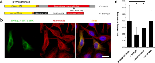
Interaction and localization of TPPP/p25 and SIRT2 in living HeLa cells as detected by immunofluorescence microscopy coupled with BiFC technology. (a) Scheme of the applied BiFC constructs. (b) Co-localization of the TPPP/p25-SIRT2 complex (green) with the MT network (red). MT network was stained with Alexa546, nuclei was counterstained with DAPI (blue). Scale bar: 10 μm. (c) BiFC signal (green) of the assembly of VenusN-TPPP/p25 and VenusC-SIRT2 and the effect of unlabelled TPPP/p25, α-synuclein and MZ25 was quantified as described in the Materials and Methods. The data are presented as mean ± SD, in each case at least 90 cells were analysed. *p = 2.45E-21 when control (BiFC) cells were compared with those co-transfected with unlabelled TPPP/p25 and *p = 6.76E-06 with unlabelled α-synuclein (two-sided, unpaired Student’s t-test).

