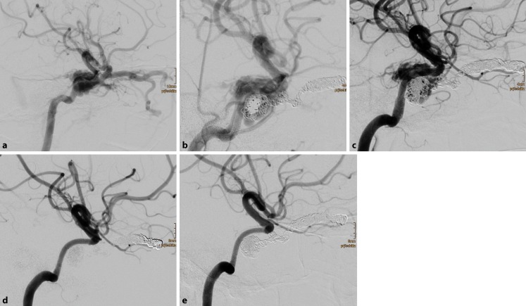
Fig. 2.
Traumatic direct carotid-cavernous sinus fistula in a 17 year-old female patient after severe head trauma (a). Chemosis, exophthalmos, and elevated intraocular pressure were due to massive arteriovenous shunt from the right internal carotid artery (ICA) into the adjacent cavernous sinus. Endovascular treatment in one session comprised insertion of 24 detachable coils with nylon fibers into the right superior ophthalmic vein and the right cavernous sinus. Thereafter, six flow diverters were deployed in the cavernous segment of the right ICA, with a significant reduction of the arteriovenous shunt (b). Early follow-up digital subtraction angiography (DSA) 6 d later revealed a residual arteriovenous shunt, which was treated with four additional flow diverters (c). Further DSA examinations 2 months (d) and 8 months (e) after the treatment showed the occlusion of the traumatic arteriovenous fistula (modified Rankin Scale 0)

