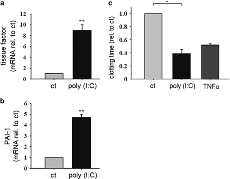
Figure 2.
Effect of poly (I:C) on the endothelial expression of procoagulatory factors and clotting time. HMEC were stimulated with poly (I:C) (10 μg/ml) for 12 h and the expression of tissue factor (a) and PAI-1 (b) was analyzed by RT–PCR (n=4, *P<0.05. mean±s.e., statistics with t-test (sigma plot); rel. to ct, relative to control). Comparable results were obtained in two series of independent experiments. (c) HMEC were stimulated with poly (I:C) (10 μg/ml) or TNFα (5 ng/ml) as a positive control for 24 h and then lysed. Whole blood samples were stimulated with cell lysates and the clotting time was analyzed as described in the Materials and methods section (n=5–6, *P<0.05. mean±s.e., statistics with t-test (sigma plot); rel. to ct, relative to control). Comparable results were obtained in two series of independent experiments. HMEC, human microvascular endothelial cell; poly (I:C), polyriboinosinic:polyribocytidylic acid; RT–PCR, reverse transcription–PCR. **P<0.01.

