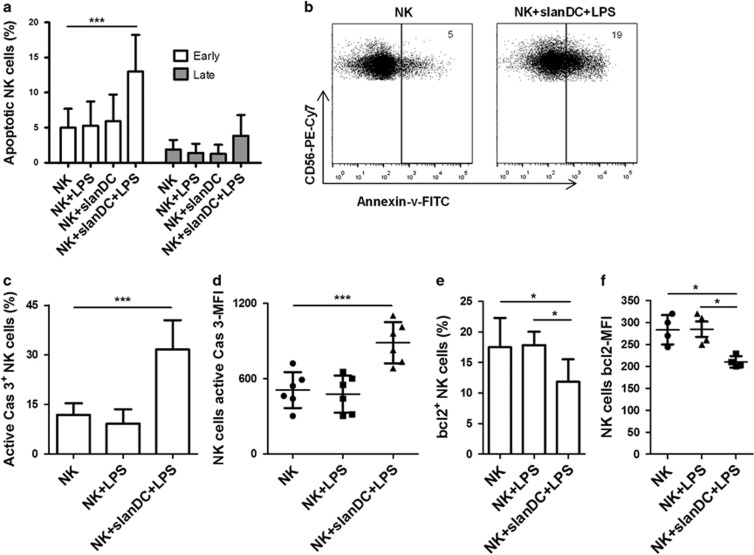
Figure 3.
Increased proportion of apoptotic NK cells after coculture with slanDCs. Freshly isolated NK cells and slanDCs were coincubated for 16 h in the presence or absence of LPS. NK cells were also cultured with or without LPS. (a) NK cells were stained for Annexin-V and 7AAD and evaluated for apoptosis using flow cytometry. The percentage of early (Annexin-V+7AAD−) and late (Annexin-V+7AAD+) apoptotic cells are shown (n=9/group). (b) NK cells were stained for Annexin-V, and dot plots are shown. The data are representative of nine independent experiments. The values represent the percentage of Annexin-V+ cells. (c) NK cells were stained for intracellular active caspase-3 expression, and the percentage of active caspase-3+ cells is shown (n=6/group). (d) NK cells were stained for intracellular active caspase-3 expression, and the MFI of active caspase-3+ cells is shown (n=6/group). (e) NK cells were stained for intracellular bcl2 expression, and the percentage of bcl2+ cells is shown (n=6/group). (f) NK cells were stained for intracellular bcl2 expression, and the MFI of bcl2+ cells is shown (n=4/group). (a, c–f) The data are shown as the mean±s.d. and are representative of four to nine independent experiments. **P<0.01 and ***P<0.001, one-way ANOVA followed by Bonferroni posttests (a, c–f).

