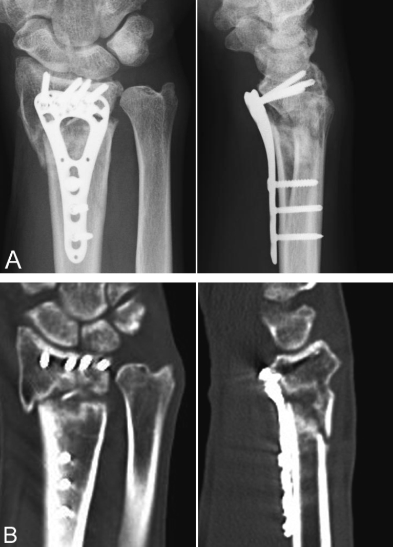
Fig. 3.
(A) Posteroanterior and lateral radiographs of the operated wrist showing a distal radius nonunion with 1.5-mm ulnar minus variance and prominence of the volar plate at the watershed line.
(B) Computed tomography scans showing segmental bone defects in the distal radius and sclerotic changes at the fracture edges.

