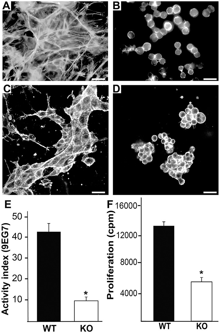
Fig. 8.

Talin 1/2 KO CD cells have major spreading, tubulogenesis and proliferation defects. (A,B) Wild-type and talin 1/2 KO CD cells were grown in 10% FBS on plastic, stained with rhodamine-phalloidin and visualized by immunofluorescent microscopy. Scale bars: 50 μm. (C,D) Wild-type and talin 1/2 KO (KO) CD cells placed in 3D collagen and Matrigel gels for 7 days in the presence of 5% FBS were stained with rhodamine-phalloidin and visualized by confocal microscopy. Scale bars: 50 μm. (E) The activation index of the β1 integrin was determined as described in the Materials and Methods. Data are mean±s.d. of three experiments performed in triplicate. *P<0.05 between wild-type and talin 1- and talin 2-null cells. (F) Thymidine incorporation in wild-type and talin 1/2 KO CD cells plated on Ln-511 in the presence of 10% FBS was measured as described in the Materials and Methods. Data are mean±s.d. of three experiments performed in triplicate. *P<0.05 (between wild-type and talin 1/2 KO cells).
