Fig. 6.
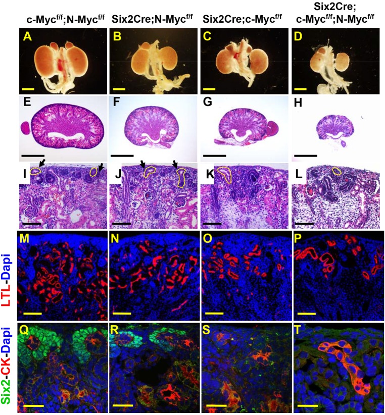
c- and N-Myc play partially redundant roles in nephron progenitor renewal and nephron endowment. Whole-mount images (A-D) or sections through P1 urogenital systems/kidneys of wild-type (c-Mycflox/flox;N-Mycflox/flox; A,E,I,M,Q), N-Myc (Six2Cre;N-Mycflox/flox; B,F,J,N,R), c-Myc (Six2Cre;c-Mycflox/flox; C,G,K,O,S) or c/N-Myc double mutant (Six2Cre;c-Mycflox/flox;N-Mycflox/flox; D,H,L,P,T) kidneys. E-L show low (E-H) or high (I-L) magnification images of H&E-stained kidneys. M-T show sectioned P1 kidneys stained with antibodies to the proximal tubule marker lotus tetragonolobus lectin (LTL, red) and DAPI (blue) (M-P) or Six2 (green), the collecting duct marker cytokeratin (CK, red) and DAPI (blue) (Q-T). Arrows indicate cap mesenchyme and ureteric bud is outlined. Scale bars: 1 mm (A-H); 100 μm (I-L); 200 µm (M-P); 30 µm (Q-T).

