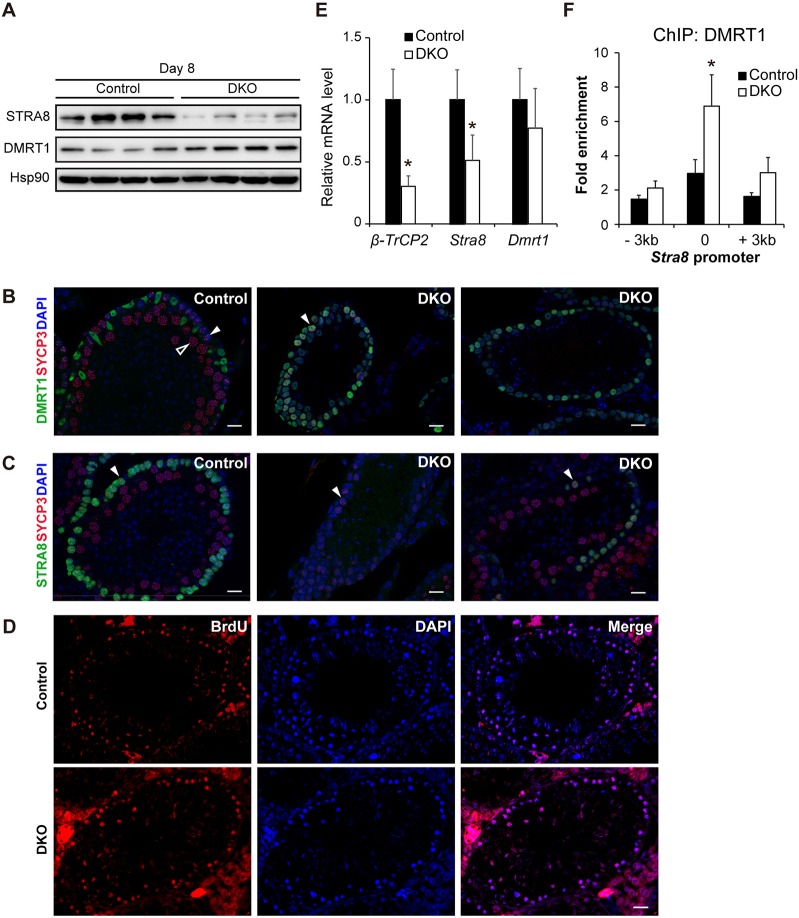
Fig. 5.
Accumulation of DMRT1 and reduction of STRA8 in β-TrCP1/2 DKO testes. (A) Immunoblot analysis of the indicated proteins in the testis of four control (Stra8-Cre, control) and four β-TrCP1/2 DKO mice at 8 dpp. Hsp90 was examined as a loading control. (B) Immunofluorescent staining of sectioned adult seminiferous tubules for DMRT1 (green) and SYCP3 (red); DAPI staining of DNA is blue. In control testes (left panel), preleptotene spermatocytes (closed arrowhead) have speckled distribution of SYCP3 and lack DMRT1, whereas pachytene spermatocytes (open arrowhead) have filamentous SYCP3 localized to synaptonemal complexes. In double-mutant tubules (middle and right panels), preleptotene spermatocytes (closed arrowhead) ectopically express DMRT1 and pachytene spermatocytes are rare. (C) Immunofluorescent staining of sectioned seminiferous tubules STRA8 (green) and SYCP3 (red); DAPI staining of DNA is blue. In control testes (left panel), STRA8 is strongly expressed in preleptotene spermatocytes (closed arrowhead), whereas in double mutant testes (middle and right panels), STRA8 is absent or weakly expressed in these cells (closed arrowheads). Scale bars: 20 μm. (D) Analysis of BrdU incorporation (red) in sections of control and β-TrCP1/2 DKO testes at 21 dpp. DAPI staining of DNA is blue. Scale bars: 20 μm. (E) RT-qPCR analysis of β-TrCP2, Stra8 and Dmrt1 mRNAs in the testis of control and β-TrCP1/2 DKO mice at 8 dpp. Data are mean±s.e.m. for four mice of each genotype. *P<0.05 versus control (unpaired Student's t-test). (F) ChIP-qPCR analysis of DMRT1 associated with the Stra8 promoter in the testis of control and β-TrCP1/2 DKO mice at 8 dpp. Regions (3 kb) upstream and downstream of the Stra8 promoter were examined as negative controls. Data are mean±s.e.m. for three mice of each genotype. *P<0.05 versus control (unpaired Student's t-test).

