Fig. 7.

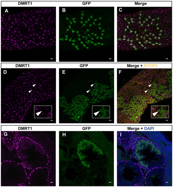
Post-translational regulation of DMRT1 in germ cells. (A-C) Immunofluorescent staining of whole-mount seminiferous tubule for DMRT1 (magenta) and GFP (green). Strongly double-positive cells at surface of tubule are spermatogonia. (D-F) Immunofluorescent staining of whole-mount seminiferous tubule DMRT1 (magenta), GFP (green) and SYCP3 (orange). Large clones of GFP-positive cells below the tubule surface are spermatocytes and are SYCP3 positive and DMRT1 negative (arrowheads indicate examples of such cells). DMRT1-positive cells interspersed among SYCP3-positive spermatocytes are spermatogonia and Sertoli cells. Insets show the enlarged images of the cells indicated by arrowheads. (G-I) Immunofluorescent staining of seminiferous tubule section for DMRT1 (magenta) and GFP (green). Cells expressing GFP and DMRT1 are round spermatids. GFP immunofluorescence in whole-mount tubules is inefficient in spermatids and hence the strongly GFP-positive germ cells in E and F are primarily spermatocytes; GFP expression in spermatids is more readily apparent in cross-section, as in H and I, where the majority of strongly GFP-positive cells are round spermatid. Scale bars: 20 μm.
