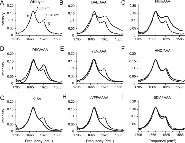
Figure 3.
FTIR spectroscopy of wild-type and alanine mutants of C55. Comparison of FTIR spectra of wild-type C55 (A) reconstituted into membrane bilayers with spectra of the C55 protein containing alanine mutations at (B) DAE, (C) FRH, (D) DSG, (E) YEV, (F) HHQ, (G) K, (H) LVFF, and (I) EDV. The bilayers were formed with dimyristoylphosphocholine (DMPC) and dimyristoylphosphoglycerol (DMPG) in a molar ratio of 10:3. The molar ratio of C55:total lipid was 1:50.

