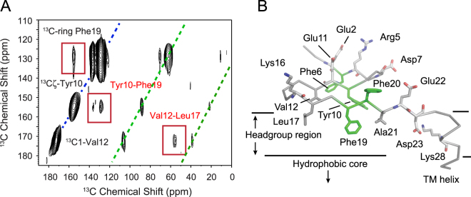
Figure 6.
Structure of the extracellular sequence of C55. (A) Solid-state DARR NMR of intra-residue contacts between Val12 - Leu17 and Tyr10 - Phe19. The C55 peptide labeled with 13Cζ-Tyr10, 13C1-Val12, 13Cα-Leu17 and 13C-ring-Phe19 was reconstituted into DMPC:DMPG bilayers as previously described18. (B) Model of the C55 extracellular region based on FTIR and NMR data showing the regions of the β-secondary structure, Val12 - Leu17 and Tyr10 - Phe19 contacts, and orientations of Phe19-Phe20 relative to the membrane bilayer surface.

