Figure 2.
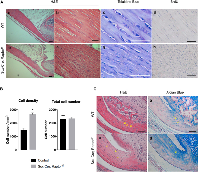
Loss of mTORC1 signaling causes abnormalities in Achilles tendons. (A) Histology of Achilles tendons from wild-type (WT) (a-d) and Scx-Cre; Raptor f/f littermates (e–h) at postnatal day 30 (P30). Low magnification (5X) image of Hematoxylin and Eosin (H&E) staining of Achilles tendon (a and e). High magnification image (40X) of H&E (b and f), toluidine blue (c and g), and BrdU (d and h) staining of the mid-substance of Achilles tendons. Scale bars in a and e indicate 200 μm, and all other scale bars indicate 50 μm. (B) Quantification of cell density and total cell number using H&E-stained sections of Achilles tendons (n = 3, *p < 0.05). (C) H&E staining of the enthesis of the Achilles tendons from WT (a) and Scx-Cre; Raptor f/f littermates (c) at P30. Alcian blue staining of the enthesis of Achilles tendons from WT (b) and Scx-Cre; Raptor f/f littermates (d) at P30. Yellow arrows indicate representative fibrocartilage cells. Scale bars indicate 100 μm.

