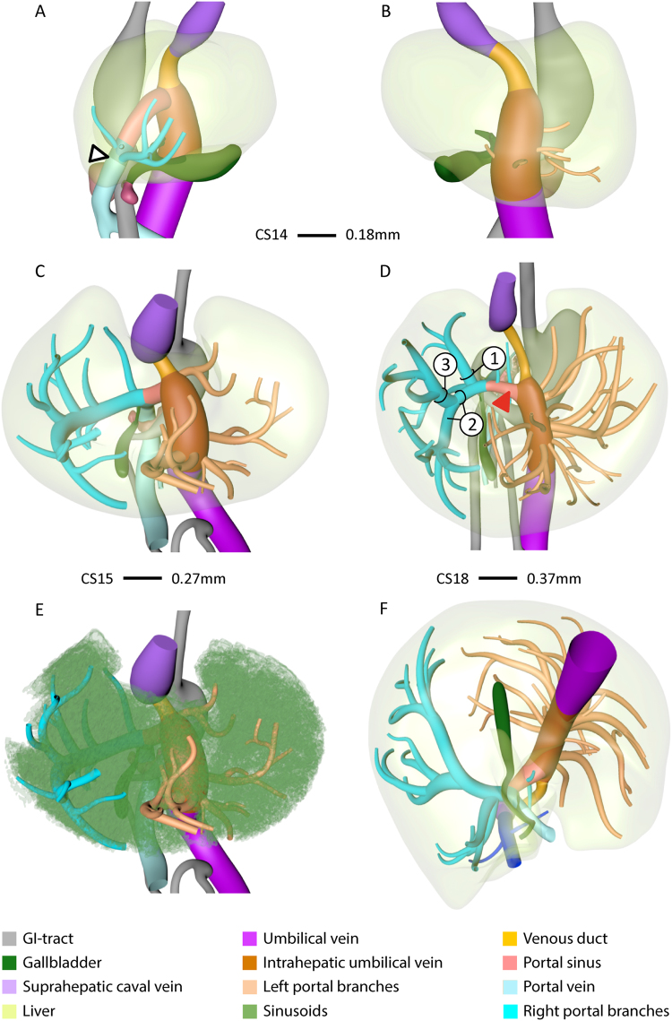
Figure 1.
The appearance of portal veins in the liver. Right-sided (A), left-sided (B), ventro-cranial (C–E) and caudal (F) views of CS14 (A,B), CS15 (C,E), and CS18 (D,F) livers. Panel E is C with sinusoids retained. Blue and peach: portal veins in right and left hemi-livers, respectively. Between CS14 and CS18, the number of large portal branches was pruned to 3 in the right hemi-liver (D) in between the sinusoidal network (E), while >10 branches remained in the left hemi-liver. Note that portal veins do not cross Cantlie’s line (F). Arrowhead (A): entrance of portal vein into the liver.

