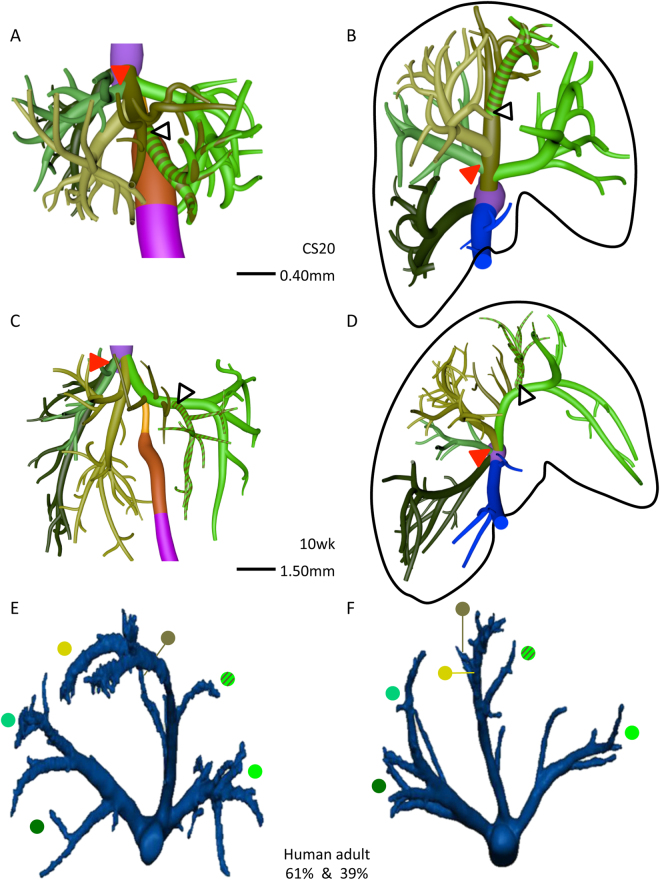
Figure 3.
The distribution of the hepatic veins in embryonic and adult human livers. Anterior (A,C) and caudal (B,D–F) views. Panels A–D show the main branches of the hepatic veins in 7 and 10 week-embryos, while panels E,F show the hepatic veins as observed by Fang c.s. (28; reproduced with permission of the publisher). The color-coded dots in panels E,F identify the hepatic veins in adults that correspond with those in embryonic livers. Each hemi-liver contains 3 hepatovenous branches in both embryos and adults. Color code is identical to that of Fig. 2.

