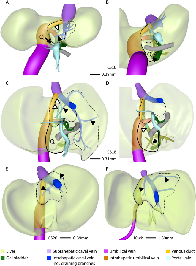
Figure 5.
Development of the caudate and quadrate lobes. Posterior (A,C,E,F) and posterio-caudal (B,D) views at CS16 (A,B), CS18 (C,D), CS20 (E) and 10 weeks (F). The caudate lobe (interrupted line) appeared at CS16 and the quadrate lobe (Q; black line) at CS18, were supplied by branches from the portal sinus (black open arrowheads), and were drained by the intrahepatic caval vein and middle hepatic vein (black arrowheads), respectively. The number and length of hepatic-vein branches that drain unto the intrahepatic caval vein (blue) varies (dotted lines; A,C,E,F) and, therefore, the boundary of the caudate process is variable.

