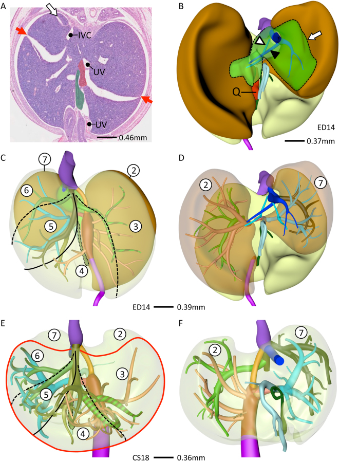
Figure 6.
Lobar distribution of veins in human and mouse livers. Cranio-ventral (C,E) and posterior (B,D,F) views. In lobated livers, both caudate lobe and process (green) are free lobes that both drain into the intrahepatic caval vein (B). The murine quadrate lobe (Q, red) is small (A,B). The left and right dorsolateral lobes (B, brown) are drained by the left lateral and right hepatic veins (D). The ventromedial lobe (E, red line) is drained by 4 main hepatic branches (left and right medial, and the two upstream tributaries of the middle hepatic vein). The distribution of the hepatic veins is similar in mouse and human livers (cf. B–D and E,F). Using the lobar boundaries Couinaud’s surgical segments can be constructed: #1: caudate; #2 and #7: dorsolateral; and ##3–6: ventromedial with 3 hepatic veins. Symbols (A): red arrows: hepatic fissures; IVC: intrahepatic caval vein, UV: umbilical vein, green: gallbladder. Color code veins is as in Figs 1 and 2.

