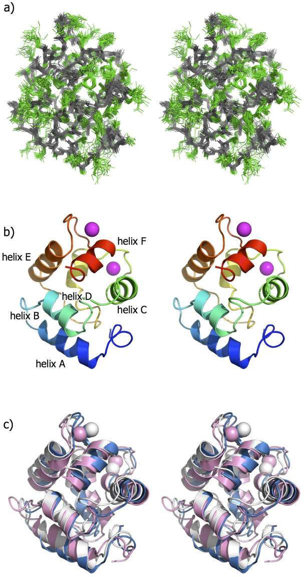
Figure 2.
NMR structures of the Pacific mackerel parvalbumin Sco j 1 in stereo. (a) Overlay of the ensemble of 20 final energy-minimized CYANA structures. The main and side chains are colored in gray and green, respectively. (b) Ribbon diagrams of the lowest energy structure. Two Ca2+ ions are shown as magenta balls. (c) Superposition of fish allergenic parvalbumin structures of Sco j 1 (white), Cyp c 1 (cyan), and Gad m 1 (pink). All the figures were drawn using PyMOL (http://www.pymol.org/).

