Fig. 5.
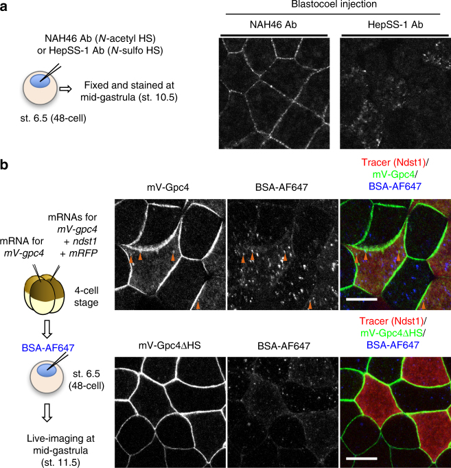
N-sulfonation-dependent internalization of the HS clusters. a Internalization assay of anti-HS antibodies. NAH46 or HepSS-1 antibody (10 ng) was injected into blastocoels at st. 6.5, and the injected embryos were fixed at st. 10.5 and stained with anti-mouse IgM secondary antibody. The animal cap region, where Wnt signaling is considered to be low10, was observed. Distribution of the injected anti-N-acetyl HS antibody showed an intercellular distribution similar to normal immunostaining of gastrula with the anti-N-acetyl HS antibody (Fig. 2e, Supplementary Fig. 3c), whereas that of the anti-N-sulfo HS antibody showed puncta inside the cells. b Facilitated internalization of the mV-Gpc4 core protein by coexpression of ndst1. Dye-labeled BSA (BSA-AF647) was injected into the blastocoel as a marker for internalized puncta. The animal cap region was observed at st. 11.5. Upper panels, in ndst1-overexpressing cells, the number of puncta of mV-Gpc4 that overlapped with BSA-AF647 (orange arrowheads) increased. Lower panels, Ndst1 did not facilitate the internalization of mV-Gpc4ΔHS, which lacks HS chains. Amounts of injected mRNA (ng/embryo): mV-gpc4, 0.067 × 2; ndst1, 0.033; mRFP, 0.33. Images are a representative of at least two independent experiments. Scale bars, 20 μm. a.u., arbitrary units

