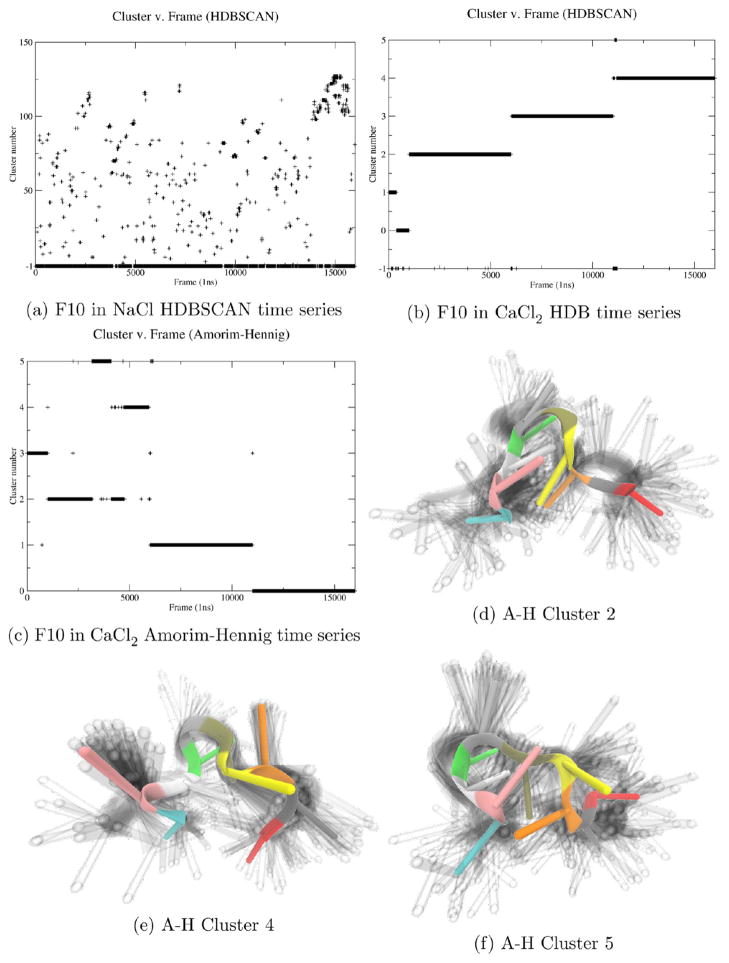
Figure 6.
HDBSCAN identified stable (a) vs unstable systems (b), and Amorim–Hennig provided finer resolution on structural changes (c–f). Visual comparison of the Amorim–Hennig clusters of the second of four trajectories (respectively beginning at frame 1, 1001, 6001, and 10 001) confirms that the method uncovered distinct conformations. The fluoridated DNA strand is colored by residue number drawn with VMD’s NewCartoon method.

