
FIGURE 4.
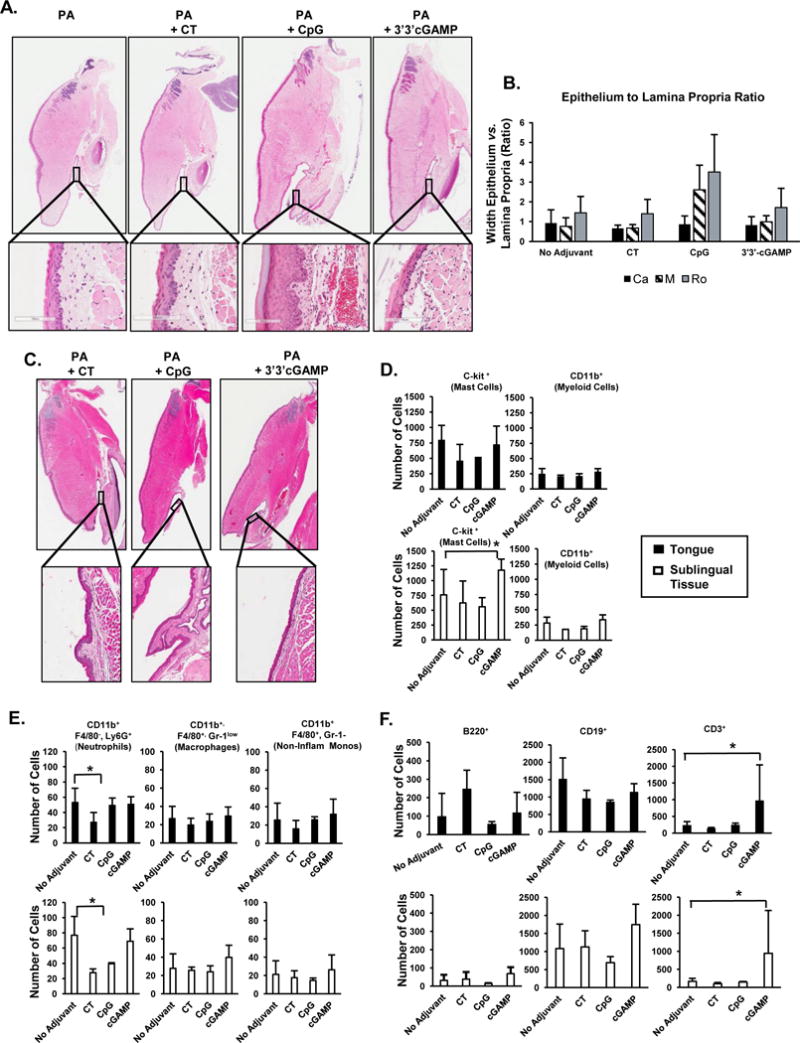
Histological structures and immune cell subsets in tongue and sublingual tissues after sublingual immunization with 3′3′-cGAMP as adjuvant. Tongue and sublingual tissues were collected 2 hours (A, B) or 42 hours (C) after a single sublingual application of PA alone, PA plus cholera toxin (CT), PA plus CpG ODN 1826 (CpG), or PA plus the STING ligand 3′3′-cGAMP (3′3′-cGAMP). Thin sections (10 μM) of paraffin-embedded tissues were stained with hematoxylin & eosin. The sublingual mucosa was divided into thirds by length and the ratio of epithelium to the lamina propria was calculated every 100 μm and averaged for each third of the sublingual tissue. Potential changes in the structure of sublingual tissues were quantified by measuring the ratio of epithelial to lamina propria width in the caudal (Ca), middle (M) and rostral (Ro) portion. (D–F) Flow cytometry analysis of immune cell subsets 2 hours after sublingual application of the vaccine formulations. (D) c-Kit+ and CD11b+ cells; (E) myeloid cell subsets; (F) B and T cells. Results are expressed a mean ± SD (n=3–6 per group). *p ≤ 0.05 compared to the PA only (no adjuvant) group (Dunnet’s test).