Fig. 2.

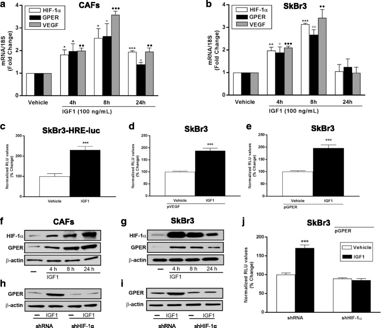
IGF1 induces the expression of HIF-1α, GPER and VEGF. mRNA expression of HIF-1α, GPER and VEGF in CAFs (a) and SKBR3 (b) cells treated for 8 hours with 100 ng/mL, as evaluated by real-time PCR. Values are normalized to the 18S expression and shown as fold changes of mRNA expression induced by IGF1 compared to cells treated with vehicle. Data are mean ± SEM of three independent experiments performed in triplicate. c Evaluation of luciferase activity in SKBR3 cells infected with a HRE reporter construct (SKBR3-HRE-luc) and treated for 18 hours with IGF1 (100 ng/mL). The luciferase activities were normalized to the protein content, evaluated in parallel plate by SRB (sulforhodamine B) assay. The transactivation of a VEGF (pVEGF) (d) and a GPER (pGPER) (e) promoter construct is observed in SKBR3 cells treated with 100 ng/mL IGF1 for 18 hours. Luciferase activity was normalized to the internal transfection control. Results are expressed as the % change of normalized RLU values relative to vehicle-treated cells. Data shown are the mean ± SEM of two independent experiments performed in triplicate. Representative immunoblots showing the increase of HIF-1α and GPER protein expression in CAFs (f) and SKBR3 cells (g) treated with 100 ng/mL IGF1 for 8 hours. The upregulation of GPER protein expression observed treating CAFs (h) and SKBR3 cells (i) for 8 hours with 100 ng/mL IGF1 is abrogated by silencing HIF-1α. β-actin serves as loading control. j The transactivation of a GPER promoter construct (pGPER) detected in SKBR3 cells treated with 100 ng/mL IGF1 for 18 hours is abrogated by HIF-1α silencing. (*), (○), p < 0.05; (**), (○○), (●●) p < 0.01; (***), (●●●) p < 0.001. CAFs cancer-associated fibroblasts, GPER G-protein estrogen receptor, HIF-1 hypoxia inducible factor-1, IGF1 insulin-like growth factor 1, VEGF vascular endothelial growth factor
