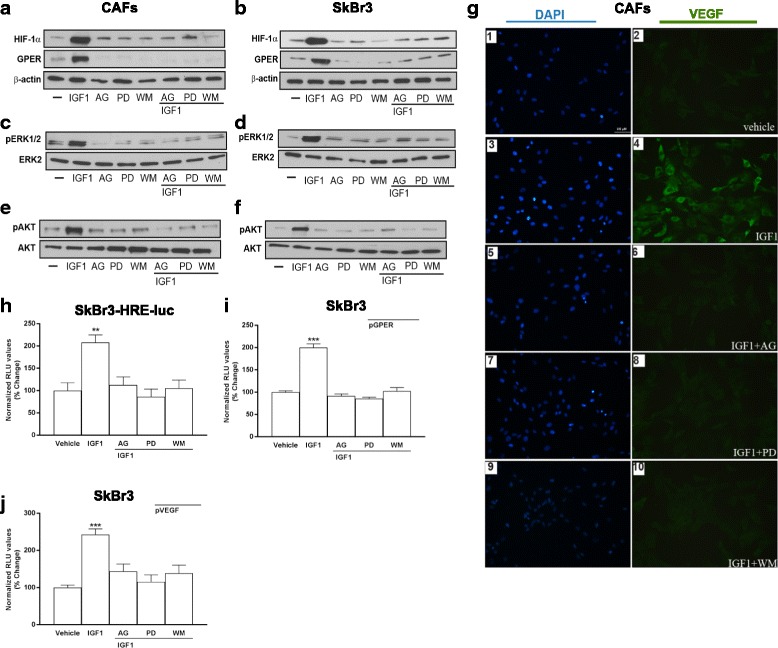
Fig. 3.

ERK1/2 and AKT signaling pathways are involved in the upregulation of VEGF expression induced by IGF1. The upregulation of HIF-1α and GPER protein expression observed treating CAFs (a) and SKBR3 cells (b) with 100 ng/mL IGF1 for 8 hours is abolished in the presence of 10 μM IGF1R inhibitor AG1024 (AG), 10 μM MEK inhibitor PD98059 (PD) and 100 nM PI3K inhibitor Wortmannin (WM). The activation of ERK1/2 and AKT (Ser 473) is prevented in CAFs (c, e) and SKBR3 cells (d, f) treated for 60 minutes with 100 ng/mL IGF1, alone and in combination with AG (10 μM), PD (10 μM) and WM (100 nM). ERK2, AKT and β-actin serve as loading control, as indicated. g VEGF protein expression in CAFs treated with 100 ng/mL IGF1 for 8 hours, alone and in combination with AG (10 μM), PD (10 μM) and WM (100 nM), as evidenced by immunfluoerscence experiment. VEGF accumulation is shown by the green signal, nuclei are stained by DAPI (blue signal), bar scale 100 μM. Results shown are representative of two independent experiments. Evaluation of luciferase activity in SKBR3 cells infected with a HRE reporter construct (SKBR3-HRE-luc) (h), and in SKBR3 cells transiently transfected with a GPER (pGPER) (i) or a VEGF (pVEGF) promoter construct (j) and treated with 100 ng/mL IGF1 for 18 h in the presence of AG, PD and WM. Luciferase activity was normalized to the internal transfection control. Results are expressed as the % change of normalized RLU values relative to vehicle-treated cells. Each data point represents the mean ± SEM of two independent experiments performed in triplicate. (**) p < 0.01; (***) p < 0.001. CAFs cancer-associated fibroblasts, GPER G-protein estrogen receptor, HIF-1 hypoxia inducible factor-1, IGF1 insulin-like growth factor 1
