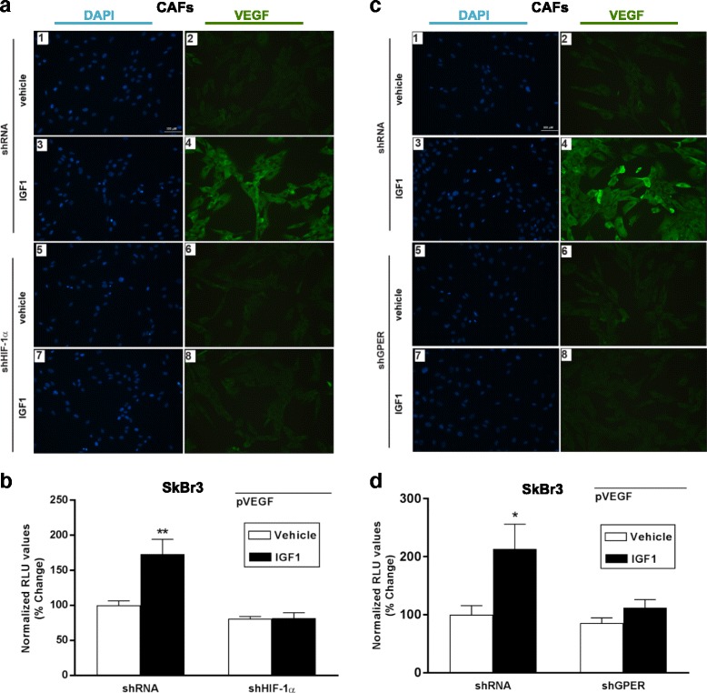
Fig. 4.

HIF-1α and GPER are involved in VEGF protein increase induced by IGF1. a Evaluation of VEGF protein expression by immunofluorescence experiment in CAFs transfected for 24 hours with control shRNA (panels 1–4) or shHIF-1α (panels 5–8) and treated with 100 ng/mL IGF1 for 8 hours, as indicated. b The transactivation of a VEGF (pVEGF) promoter plasmid observed in SKBR3 cells treated with 100 ng/mL IGF1 for 18 hours is abrogated silencing the expression of HIF-1α. c Evaluation of VEGF protein expression by immunofluorescence experiment in CAFs transfected for 24 hours with control shRNA (panels 1–4) or shGPER (panels 5–8) and treated with 100 ng/mL IGF1 for 8 hours, as indicated. In immunofluorescence experiments, VEGF accumulation is evidenced by the green signal, nuclei are stained by DAPI (blue signal), bar scale 100 μM. Images shown are representative of two independent experiments. d The transactivation of a VEGF (pVEGF) promoter plasmid observed in SKBR3 cells treated with 100 ng/mL IGF1 for 18 hours is abrogated silencing the expression of GPER. In luciferase assays, luciferase activity was normalized to the internal transfection control. Results are expressed as the % change of normalized RLU values relative to vehicle-treated cells. Each data point represents the mean ± SEM of two independent experiments performed in triplicate. (*) p < 0.05, p < 0.01 (**). CAFs cancer-associated fibroblasts, GPER G-protein estrogen receptor, HIF-1 hypoxia inducible factor-1, IGF1 insulin-like growth factor 1, VEGF vascular endothelial growth factor
