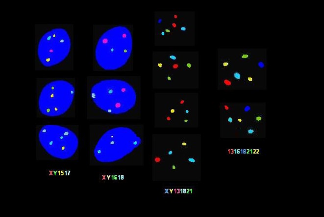
Figure 1.

Fluorescent in-situ hybridization (FISH) analysis of ejaculated human spermatozoa. FISH analysis was carried out using 4 different probe sets. In the 2 columns on the left, sperm chromatin stained with 4′,6-diamino-2-phenylindole (DAPI) appears in blue. As indicated from left to right: spermatozoa were assessed by probe sets for chromosomes X/Y/15/17, X/Y/16/18, X/Y/13/18/21 and 13/16/18/21/22 in various colors. As depicted in each cell, disomy is indicated by the appearance of multiple fluorescent signals in the same color. Spermatozoa exhibiting various occurrences of gonosomal and autosomal disomy are shown.

 This work is licensed under a