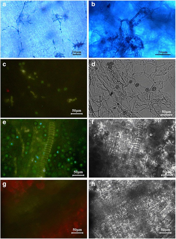
Fig. 5.

The distribution and morphology of U. esculenta in Jiaobai tissues after inoculation. a and b The fungal morphology in grey Jiaobai (a) and white Jiaobai (b). The tissues were collected from stem tips of the plants in the fields when they had five leaves, then sliced and stained with aniline blue. Images were taken by light microscope. Bars indicated 20 μm. c-f Images were taken by fluorescence microscope. Bars indicated 50 μm. Recombinant strains containing the EGFP reporter gene (UeT14::EGFP-NLS and UeT55::EGFP-NLS) were applied to inoculate. c and d Samples were collected from the new shoots, 1 month after the inoculation assays. Few spores and hyphae were observed in samples generated by tissue slice. e and f Samples were collected from the swollen stem of the inoculated plants, nearly 3 month after the inoculation assays. Full of teliospores and hyphae were detected in samples generated by tissue slice. g and h Samples were collected from wild Jiaobai (the controls) 3 month after the inoculation assays. No fluorescent signal of fungus was detected in samples generated by tissue slice
