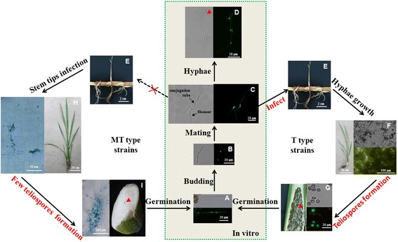
Fig. 7.

The life cycle of T and MT type strains. a-d The progress of teliospore germination (a), haploid strain stage (b), conjugation tubes formation and mating (c), dikaryotic filamentous stage (d). Bars indicated 20 μm. The red arrowhead pointed out the vacancy always appeared. e The rhizomes of Jiaobai. Bars indicated 2 cm. f and g The plant inoculated with T type strains. f The hyphae and teliospores obviously observed in inoculated plant stem shoots after 1 month. Bars were 20 cm (left) and 100 μm (right). g Grey Jiaobai formed (left) and the micromorphology of teliospore inside. Bars were 20 μm. The microscopic images (a-g) were taken by a fluorescent microscope. h The hyphae growth in white Jiaobai were observed obviously more than 1 month. Bars were 50 μm (left) and 20 cm (right). i The swollen stem of white Jiaobai with fewer scattered dark teliospore (right) and the hyphae aggregated inside (left). Bars were 100 μm. h and g The tissues were stained in aniline blue and microscopic images were taken by light microscope
