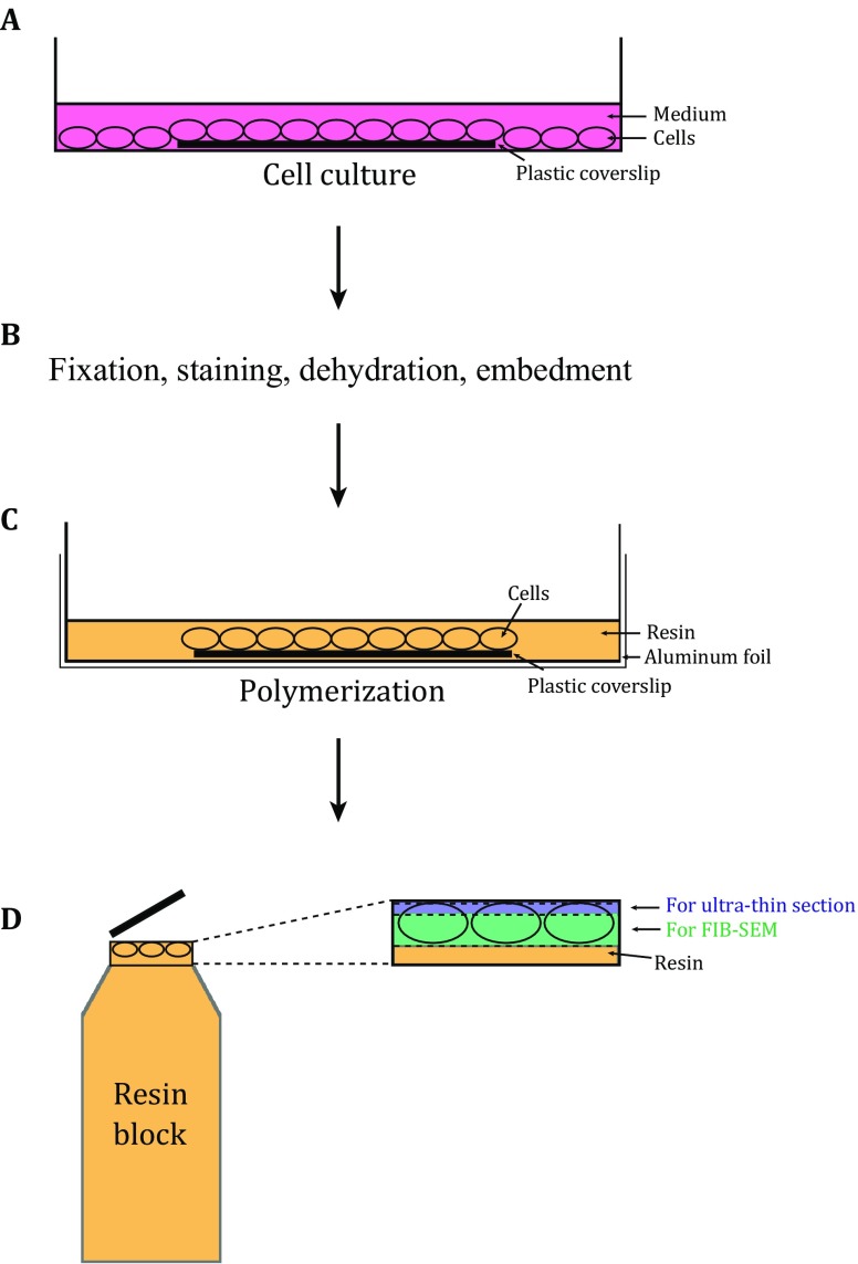
Fig. 1.
The scheme for APEX2–FIB-SEM. A The cells are cultured on sterile plastic coverslips in dishes, and transfected with the target genes fused with APEX2. The cells on the plastic coverslip are used for further EM sample preparation. B The cells on the plastic coverslip undergo fixation, DAB staining, dehydration, and resin embedment. C Before polymerization, the plastic coverslip with cells is transferred to the dish placed with aluminum foil. The cell side of the plastic coverslip is facing up. Fresh resin with accelerator is added to cover the cells. D After polymerization, the block is separated from aluminum foil and plastic coverslip, and the areas with staining patterns are cut off and stuck on the tip of an empty resin block. In order to reserve enough sample for FIB-SEM (colored with green), only three to five slices (less than 80 nm) are sectioned from the block (colored with blue) for TEM screening

