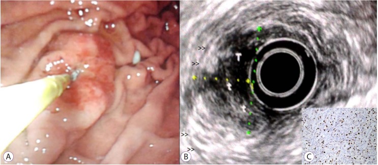
Fig. 3.
Example of a type 3 gastric neuroendocrine tumor in distal gastric body. (A) Endoscopically the lesion is quite large over 2.5 cm and is sessile with a broad, fixed base and a central depressed region. (B) At endoscopic ultrasound, the lesion can be seen to extend to touch the deep muscle layer (double arrow heads) and was predicted uT2 (N0) but after surgical resection the final pathological stage was pT2N1 (one single small node involved) with a Ki-67 of 30% (insert, C, ×200).

