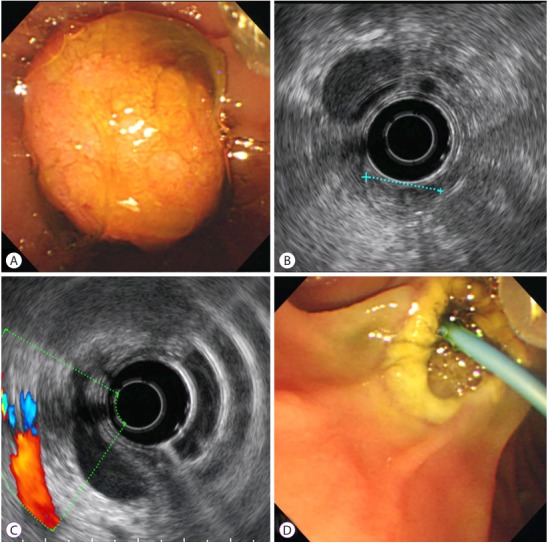
Fig. 2.

A 2-cm adenoma-like protruding lesion was found in the ampulla of Vater on endoscopy (A) and endoscopic ultrasound showed an approximately 17-mm well-defined isoechoic homogeneous mass without bile duct or pancreatic duct invasion. A dilated common bile duct is visible (B, C). After mass resection using a snare and the insertion of a biliary plastic stent, no specific complications, such as severe bleeding or perforation, were observed (D)
