
Figure 1.
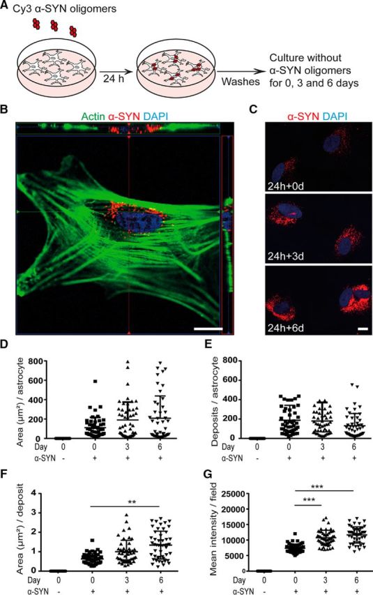
Intracellular α-SYN is stored rather than degraded. Human ESC-derived astrocytes (see Fig. 1-1A) were exposed to 0.5 μm Cy3-labeled α-SYN oligomers (see Fig. 1-1B) for 24 h, washed thoroughly, and cultured for an additional 0, 3, or 6 d in α-SYN-free medium before fixation (A). The intracellular location of the α-SYN-Cy3 after ingestion (see Fig. 1-2A) was confirmed with confocal imaging (B). Quantification of the α-SYN-Cy3 signal at days 0, 3, and 6 (C and Fig. 1-2B) reveled the total area of the α-SYN deposits per astrocyte in square millimeters (D), the number of α-SYN deposits per astrocyte (E), the mean area of the deposits in square millimeters (F), and the mean intensity of the intracellular α-SYN-Cy3 per field (G). Western blot analysis confirmed the presence of high-molecular-weight α-SYN species in the astrocytes at all three time points (Fig. 1-2C), demonstrating that only minimal degradation of the ingested oligomeric α-SYN occurred, whereas the monomeric α-SYN was completely degraded during the 6 d time period (see Fig. 1-3A, B). Scale bars: B, 10 μm; C, 20 μm. Data are presented as mean ± SD from four independent experiments and the levels of significance were set to *p < 0.05, **p < 0.01, and ***p < 0.001 (E–G).