
Figure 5.
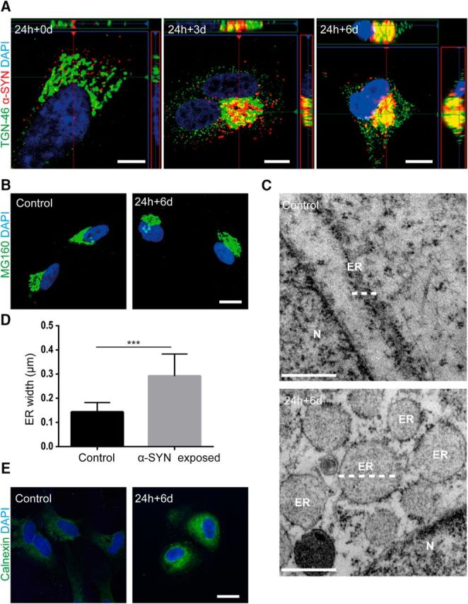
Intracellular storage of α-SYN in the TGN region causes ER swelling. Confocal microscopy imaging showed that α-SYN-Cy3 inclusions localized to the region of the TGN46+ TGN over time (A and Fig. 5-1A). Immunostainings with specific anti-Golgi complex antibodies demonstrated that the accumulation of ingested α-SYN oligomers in the TGN region did not induce Golgi fragmentation (B). TEM analysis of astrocytes 6 d after α-SYN oligomer exposure (24 h + 6 d) indicated that the α-SYN storage induced ER swelling (C) (white dotted lines indicate ER). Quantification of the ER width confirmed the induced ER swelling in oligomer-exposed astrocytes (D). Representative images from immunostainings with the anti-calnexin antibody showed a higher expression of calnexin in the perinuclear region of α-SYN oligomer-exposed astrocytes compared with control (E and Fig. 5-1B). Scale bars: A, 10 μm; B, 20 μm; C, 500 nm; E, 20 μm. Data are presented as mean ± SD from 14 α-SYN-exposed and 13 control astrocytes and the levels of significance were set to *p < 0.05, **p < 0.01, and ***p < 0.001 (D).